The extraordinary case of the ferocious female moles
Their genital anatomy, musculature and aggressiveness have made them a model for studying the phenomenon of female masculinization — and demonstrate that sometimes, it’s not easy to tell the difference between male and female
Support sound science and smart stories
Help us make scientific knowledge accessible to all
Donate today
Rafael Jiménez Medina learned how to hunt elusive Iberian moles in the fields of southern Spain in the 1980s, when he was a young PhD student in genetics at the University of Granada.
A local hunter of the moles (Talpa occidentalis) taught him how to capture these solitary, aggressive and territorial animals. The moles dig subterranean galleries and labyrinths in the meadows of the Iberian Peninsula, especially those with soft soils rich in earthworms, their favorite food. Such activity can benefit the soil — by aerating or mixing it — but the moles’ presence and constant movement in cultivated land raise the ire of farmers, who pay hunters to get rid of them.
Jiménez Medina had a different motivation for hunting these subterranean mammals. His doctoral project was to visualize and analyze their chromosomes, which meant collecting, preparing and examining samples from the testes of males. His lab analyses led to a curious finding: Some of the moles he had identified as males were in fact genetically females — that is, their sex chromosomes were XX (female) and not XY (male).
The confusion, we now know, stems from the unusual composition of the reproductive organs of female moles. In contrast to most female mammals, which have only ovaries, female Iberian moles also have testicular tissue. This tissue anatomically resembles male testicles but differs in that it produces testosterone but no sperm. The female mole’s organs are composed of both an ovarian and a testicular portion and are known as ovotestes.
In addition, female moles have a clitoris covered with a foreskin and with an elongated appearance that resembles a penis; they urinate through this structure. Another unique anatomical feature is that during these females’ juvenile stage, the vaginal orifice remains closed.
So fascinating were his findings that Jiménez Medina, after finalizing his doctoral studies, set aside his work in chromosomal analysis to immerse himself in the study of the enigmatic sexual development of these animals. His work, and that of other colleagues over the past decades, would turn this small mammal species — each of which could fit in the palm of your hand — into a model for the study of female masculinization and the biological mechanisms that underlie it.
These moles and other species whose females exhibit typically male traits — such as the spotted hyena — “have become the paradigms of female masculinization,” Jiménez Medina and colleagues wrote in the 2023 Annual Review of Animal Biosciences, wherein they detail the rewards and challenges of studying the creatures’ anatomy and behavior. These exceptional cases invite us “to reconsider the concepts of femininity and masculinity” in the field of reproductive biology, the authors write.
Female Iberian moles show intersex traits both inside and out. Here, a photo of the external genitalia of a female reveals the anus and the upright clitoris covered by a foreskin.
The presence of both male and female sexual characteristics in a single creature has been documented in many invertebrate and fish species, but in mammals this phenomenon is mainly restricted to isolated cases — to reports of individuals rather than a whole species. Such anatomical amalgamation has been described in humans for centuries: As far back as ancient Greece, surgical interventions on patients with ambiguous genitalia were reported.
Formerly called hermaphroditism (in reference to Hermaphrodite, a character from Greek mythology with voluptuous breasts and male external genitalia), the characteristics are today referred to as intersex, explains molecular geneticist Francisca Martínez Real of the Max Planck Institute for Molecular Genetics in Berlin.
What’s remarkable about the moles, however, is that this intersexuality doesn’t occur just in a few individuals: it is the norm in females. And it is not exclusive to Iberian moles: Intersexuality has been identified in eight mole species and in a species related to moles called desmans, which live near aquatic environments such as rivers and lakes. These male characteristics provide these females with tools to survive in extreme environments.
The power of testosterone
A key consequence of female moles having ovotestes is their high levels of testosterone, the major sex hormone in males (essential to the development of male growth and masculine characteristics), which directly influences the female moles’ anatomy and behavior.
But their testosterone production is seasonal, varying as the size of testicular tissue in the ovotestes changes dramatically throughout the year, explains Martínez Real, who began studying the regulation of sexual development in mammals in Jiménez Medina’s laboratory in the early 2000s. At the end of the fall, during the winter and in the beginning of spring, the ground fills up with earthworms and life becomes easier for the moles. At this time, the testicular tissue shrinks and, therefore, the females are less aggressive. Between September and May — and most intensely between November and March — the females allow the males to enter their territory for mating, says Jiménez Medina. Outside the mating period, interactions between the two sexes are reduced mainly to territorial confrontations.
All female Iberian moles have ovotestes: gonads — reproductive glands that produce gametes — containing both ovarian and testicular tissue. Ovotestes are located where simple ovaries are found in other species. These females make eggs in the ovarian part of their ovotestes, while the testicular portion secretes testosterone but makes no sperm.
During the summer months, things get more complicated. In this time of dry, hard ground and scarce food, it is more difficult for the animals to dig tunnels and galleries, and more strength is needed. The testicular part of the ovotestes enlarges, and testosterone blood levels in females averages 2.62 nanograms per milliliter in adults and 5.5 ng/ml in juveniles. These figures are in the same order of magnitude as those of males at the same period: 4.9 ng/ml in adults and 3.9 ng/ml in juveniles. By way of comparison, an adult female human has testosterone levels of about 0.5 ng/ml, equivalent to one-tenth of those of an average adult male, at about 5 ng/ml of testosterone.
Exactly how these seasonal variations in testosterone production occur in females remains a question that Jiménez Medina and his colleagues are still investigating. What is clear is that the higher testosterone levels give female moles the muscularity necessary to achieve key tasks: building galleries, making hunting grounds to catch earthworms, defending their territory. “The musculature they have is impressive,” says Jiménez Medina. When you dissect a mole — whether female or male — what you see are the muscles of a bodybuilder, he says.
“Testosterone is linked to aggression in all kinds of mammals and other vertebrates,” says Jenny Graves, an evolutionary geneticist at La Trobe University in Australia who has studied sex determination in mammals. Although in most mammal species the male is the biggest, strongest and most aggressive, she says it is very interesting to see examples like moles, in which females are the ones protecting their territory.
Genome rearrangements
In mammals, sex is genetically determined, so the intersexuality of mole females must have a genetic basis. In search of clues, Martínez Real and Jiménez Medina were part of a team that analyzed the genomes of the Iberian mole and one of its relatives, the star-nosed mole (Condylura cristata), whose females also develop ovotestes.
The team found that the genomes of both species have alterations that affect the activity of key genes involved in testosterone production and testis development, modifying when and how much the genes are turned on or off in the sex organs of females, as reported in Science in 2020.
One of these alterations affects the activity of a gene called CYP17A1, which encodes an enzyme that controls the production of male sex hormones, including testosterone. Iberian and star-nosed moles have three copies of this gene, while most mammals have only one. But the two extra copies of the gene do not function, and the important change lies in a modified copy of a nearby DNA sequence that regulates gene activity. This modification in the regulatory elements increases CYP17A1 activity in both the testes of males and in the testicular tissue of the ovotestes of females, resulting in increased testosterone production in both sexes.
Both the Iberian mole and the star-nosed mole have a genetic alteration (left) that ups the activity of the gene, CYP17A1, involved in controlling the production of male sex hormones such as testosterone. That translates into a boost of CYP17A1 activity in the reproductive organs of the moles, especially in the female testicular tissue, compared with that of mice (right). This and other genetic shifts may help explain why the female moles show traits and behavior common in males of other mammals, which have only one copy of CYP17A1.
In addition, in mammals there is another gene, CYP19A1, that is commonly active only in the ovaries and which converts testosterone into female hormones. This gene could reverse the effects of increased testosterone production from the changes in the CYP17A1 gene. But in moles, CYP19A1 is activated only in the ovarian part of the ovotestes, not in the testicular part. That means that it doesn’t interfere with testosterone production.
A third alteration affects the activity of yet another gene, known as FGF9, which contributes to the development of the testes in mammals. Both the Iberian mole and the star-nosed mole have a piece of DNA inverted in a region that’s key to the regulation of this gene. As a result, Martínez Real and colleagues found, the gene is active in the reproductive organs of females during embryonic development, when in other mammals it is active only in the males. Its activity delays ovarian development and favors the development of the testicular tissue of what will later become ovotestes, explains Martínez Real.
Experiments that introduced such genetic alterations in mice showed that they play a role in the development of females with masculinized traits. These mice — males and females — received an extra copy of the sequence that regulates CYP17A1 activity and produced greater amounts of testosterone: Females produced twice as much as females whose genomes were not altered, and males produced three times more than unaltered males. Meanwhile, female mice engineered to increase the activity of the FGF9 gene, similar to its increase in the moles, developed testes.
Different ways of being female
Do the presence of ovotestes, large muscles and aggressive behavior make these female moles less “feminine”? Jiménez Medina says the answer is no. “From a biological point of view, the most feminine thing is to become a mother,” he says, since anatomical female-male sexual forms exist for the purpose of sexual reproduction. And female moles carry out this role with no problems: They have about four pups a year, which they care for and nurse for about a month. “They build a new nest specially fitted out to give birth and nurse their young,” says Jiménez Medina, and they do so using layers of dry leaves that probably serve to ensure waterproofing and thermal insulation.

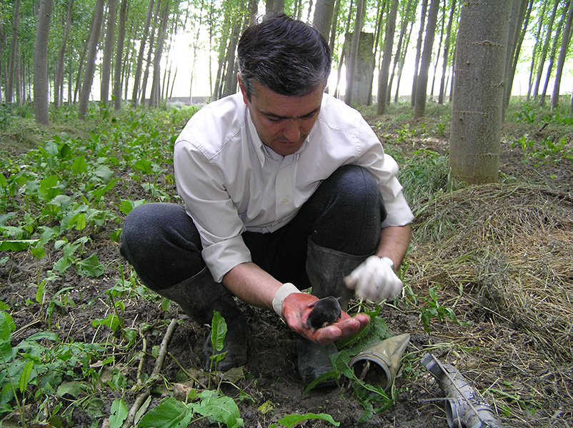
Developmental geneticist Rafael Jimenez Medina holds a recently captured mole in a field in Spain.
CREDIT: PHOTO COURTESY OF RAFAEL JIMÉNEZ MEDINA
The female moles are also very efficient nursers. Their pups can increase their birth weight by as much as 45 grams after one month of lactation, perhaps reflecting the high nutritional quality of the mother’s milk. Females with an average weight of 53.5 grams can produce litters averaging 144.12 grams at weaning, suggesting a higher reproductive effort than other female mammals — specifically, several species of shrews — with which they have been compared.
In biological terms, the female moles make it clear that there is no single way to be female and, in exceptional cases, the development of masculinized traits could be related to adaptation to a challenging environment, such as living underground.
Kay Holekamp, a behavioral ecologist at Michigan State University who has been studying female spotted hyenas for decades, says that they and female moles have a strange mix of traits. But to say that these females are masculinized “is only a fraction of the story,” she adds: There are aspects of their nervous system and behavioral repertoires that are not masculinized at all and are, in fact, “very feminine.”
“The interesting thing about these animals,” she says, “is that they are, ultimately, chimeras.”
Article translated by Debbie Ponchner
10.1146/knowable-071423-1
TAKE A DEEPER DIVE | Explore Related Scholarly Articles


